Kód: 06620871
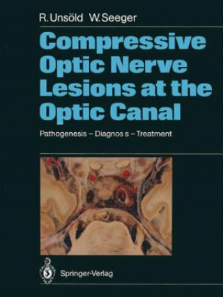
Compressive Optic Nerve Lesions at the Optic Canal
Autor Wolfgang Seeger
The optic canal, in particular its intracranial end, represents a "locus minoris resistentiae" for optic nerve compression in a variety of pathologic conditions. The intracranial optic nerve shares the limited space within this na ... celý popis
- Jazyk:
 Angličtina
 - Väzba: Brožovaná
- Počet strán: 140
Nakladateľ: Springer-Verlag Berlin and Heidelberg GmbH & Co. KG, 2011
- Viac informácií o knihe

123.03 €

Skladom u dodávateľa v malom množstve
Odosielame za 10 - 15 dní
Potrebujete viac kusov?Ak máte záujem o viac kusov, preverte, prosím, najprv dostupnosť titulu na našej zákazníckej podpore.
Pridať medzi želanie
Mohlo by sa vám tiež páčiť
-

Writing Systems
1337.06 € -

Marvel Book
19.56 € -24 % -

Asian Green
21.81 € -22 % -

Far Cry: Absolution
9.41 € -28 % -

Danganronpa: The Animation Volume 2
12.80 € -17 % -

Tales of Ise
12.90 € -30 % -

Star Wars: Thrawn Ascendancy (Book I: Chaos Rising)
11.46 € -18 % -

Mesoscale Convective Systems over the Tropics
89.74 € -

Learning ICT with Maths
58.90 € -

Vector Analysis and Cartesian Tensors, Third edition
130.61 € -

Fundamentals of Plasticity in Geomechanics
130.61 € -

Bridges and Barriers
53.78 € -

Essential Maths 7S Homework Answers
4.60 € -

Communicate Like a Leader
24.99 € -

Law and Development of Middle-Income Countries
43.53 € -15 % -

Adaptive Control of Solar Energy Collector Systems
123.03 € -

Design of Shape Memory Alloy (SMA) Actuators
78.98 € -

Coloriages mystères Les grands classiques Disney
21.20 € -

Dziewczyna, która skoczyła do morza. Mężczyzna z honorem. Tom 3
7.57 € -30 % -

Jane Eyre
14.44 € -

Trezwak
41.07 € -9 %
Darujte túto knihu ešte dnes
- Objednajte knihu a vyberte Zaslať ako darček.
- Obratom obdržíte darovací poukaz na knihu, ktorý môžete ihneď odovzdať obdarovanému.
- Knihu zašleme na adresu obdarovaného, o nič sa nestaráte.
Viac informácií o knihe Compressive Optic Nerve Lesions at the Optic Canal
Nákupom získate 304 bodov
 Anotácia knihy
The optic canal, in particular its intracranial end, represents a "locus minoris resistentiae" for optic nerve compression in a variety of pathologic conditions. The intracranial optic nerve shares the limited space within this narrow passage with the carotid and ophthalmic artery, all being surrounded by bone and rigid dura. Any pathological condition going along with an increase of soft tissue volume, such as in optic nerve sheath tumors, parasellar neoplasms, dolichoectasia of the carotid and/ or ophthalmic artery, hematomas, etc. , or reduction of the lumen of the bony optic canal by hyperpneumatization of the sphenoid sinus, hyperostosis or developmental abnormalities must act as a space-occupying lesion causing optic nerve compression either by pressing the nerve against the vessel or the neighboring dura or bone. The spectrum of clinical signs and symptoms of optic nerve compression in this area is rather wide and includes acute as well as slowly progressive visual loss and all kinds of visual field defects in the presence of a normal disk, papilledema, pri mary optic atrophy or cavernous optic atrophy mimicking var ious clinical disease entities such as retrobulbar optic neuritis, anterior and posterior ischemic optic neuropathy, soft glaucoma and others. Some of the lesions causing optic nerve compression in this area are rather small and need to be visualized or excluded by thin section CT such as pneumosinus dilatans of the sphenoid bone, dolichoectasia of the internal carotid artery, small men ingiomas around the optic foramen and others.
 Parametre knihy
Zaradenie knihy Knihy po anglicky Medicine Other branches of medicine Medical imaging
123.03 €
- Celý názov: Compressive Optic Nerve Lesions at the Optic Canal
- Podnázov: Pathogenesis - Diagnosis - Treatment
- Autor: Wolfgang Seeger
- Jazyk:
 Angličtina
 - Väzba: Brožovaná
- Počet strán: 140
- EAN: 9783642733840
- ISBN: 9783642733840
- ID: 06620871
- Nakladateľ: Springer-Verlag Berlin and Heidelberg GmbH & Co. KG
- Hmotnosť: 400 g
- Rozmery: 280 × 210 × 9 mm
- Dátum vydania: 06. December 2011
Obľúbené z iného súdka
-

Diagnostic Ultrasound: Head and Neck
268.82 € -

Atlas of Ultrasound Guided Musculoskeletal Injections
97.32 € -4 % -
Gynaecological Ultrasound Scanning
75.50 € -4 % -

EACVI Echo Handbook
60.43 € -

Learning Radiology
57.16 € -1 % -

Top 3 Differentials in Radiology
115.14 € -

Chest X-Ray
24.89 € -10 % -

Webb, Muller and Naidich's High-Resolution CT of the Lung
244.33 € -7 % -

MR Imaging of the Abdomen and Pelvis
185.63 € -

Obstetric Imaging: Fetal Diagnosis and Care
362.76 € -

Diagnostic Ultrasound, 2-Volume Set
379.36 € -5 % -

Breast Imaging
89.84 € -5 % -

Fundamentals of High-Resolution Lung CT
104.39 € -10 % -

Atlas Of NEURORADIOLOGY
19.86 € -

Regional Anaesthesia, Stimulation, and Ultrasound Techniques
100.19 € -

Diagnostic Imaging: Musculoskeletal Non-Traumatic Disease
374.95 € -

MR Neuroimaging
210.83 € -2 % -

Breast Ultrasound
158.68 € -

Radiology of the Post Surgical Abdomen
136.55 € -

Essentials of Radiology
90.35 € -4 % -

Musculoskeletal Imaging: The Essentials
150.90 € -

Essentials of Pediatric Radiology
182.45 € -

MRI of the Fetal Brain
114.02 € -

Pediatric Radiology: The Requisites
135.53 € -

Practical Manual of Echocardiography in the Urgent Setting
76.62 € -

Magnetic Resonance Cholangiopancreatography (MRCP)
145.06 € -

Pocket Atlas of Sectional Anatomy, Volume 3: Spine, Extremities, Joints
49.17 € -1 % -
Accident and Emergency Radiology: A Survival Guide
71.50 € -

Abdominal Ultrasound
67.30 € -

Brant and Helms' Fundamentals of Diagnostic Radiology
311.85 € -

Normal Findings in CT and MRI, A1, print
19.46 € -4 % -

Neonatal Cranial Ultrasonography
124.36 € -8 % -

Ultrasound: The Requisites
135.53 € -

Osborn's Brain
362.15 € -1 % -

Hand Bone Age
78.98 € -

Callen's Ultrasonography in Obstetrics and Gynecology
127.33 € -9 % -

Echo Made Easy
37.59 € -12 % -

Neuroradiology: The Requisites
91.17 € -5 % -

Thieme Clinical Companions Ultrasound
28.88 € -4 % -
Fundamentals of Musculoskeletal Ultrasound
91.27 € -6 % -

Brant and Helms' Fundamentals of Diagnostic Radiology
234.50 € -14 % -

EACVI Textbook of Echocardiography
198.64 € -

Fundamentals of Body CT
112.17 € -

First Trimester Ultrasound Diagnosis of Fetal Abnormalities
124.16 € -2 % -
Pocket Anatomy & Protocols for Abdominal Ultrasound
69.55 € -

Perioperative Two-Dimensional Transesophageal Echocardiography
129.59 € -4 % -

Ultrasound of the Thyroid and Parathyroid Glands
132.05 € -

Analyzing Neural Time Series Data
78.77 € -16 % -
Introduction to Vascular Ultrasonography
140.55 € -5 %
Osobný odber Bratislava a 2642 dalších
Copyright ©2008-24 najlacnejsie-knihy.sk Všetky práva vyhradenéSúkromieCookies


 21 miliónov titulov
 Vrátenie do mesiaca
 02/210 210 99 (8-15.30h)